Morgellons Artifacts Found in Rainwater – Part 1
Morgellons Fibers Are Lighter Than Air
Several indicators lead me to suspect that the Morgellons filaments are in the air, inside and outside and also in rain water. I have microscopically photographed Morgellons filaments in my regular house dust, these filaments are known to physically come off of and out of some people with this condition. From what I am observing in my environment, I am suspecting that these fibers are lighter than air.
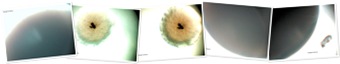
After it had been raining for some time, I collected a sample of rainwater, a small metal container was placed outside to microscopically observe what is immediately present on a slide and when rain water is cultured in agar. Below is my Environmental Study 1 experiment and my conclusions from it at 100x:
Environmental Study 1 – Day 1
4/14/2010
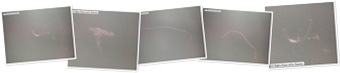
A piece of plant material also ended up in the container:
And some ‘others’ that resemble particles found in regular tap water:
This above is what is seen immediately when viewing rain water on a slide, this experiment is easily repeated by anywhere in the world. At this point the rain water was cultured in potato dextrose agar, I inserted the plant material in the dish, I wanted to see the effects of what is in the environment on plant life and has no bearing on the outcome on the goals of this experiment, as you will see.
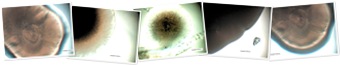
Environmental Study 1 – Day 2
The Petri Dish was taped shut, placed right side up and immediately placed inside a sealed plastic box. The dish has not been jarred to cause any contents from the base agar to be transmitted to the lid. I am studying the evaporation process, to confirm that the fibers are lighter than air – these are the condensation beads on the inside part of the lid of the dish:
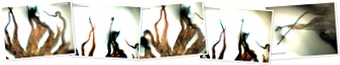
Filaments have risen from the base agar to the lid, indicating they are lighter than air. The Petri Dish was not opened at this time, these are photos of the inside lid only.
I’m noticing basically 3 different filament colors, one is a darker blue color, the other is a pinkish color and the other is translucent. I’m noticing that, at times, the blue and pink are seen together, the pink one is seen laying across the blue one. The translucent one seems to stand alone, mostly. The filaments are piercing the water bubbles, there appears to be quorum sensing prior to piercing. A pink filament is seen ‘folding’.
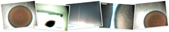
Below is a 3 series of photos to show the piercing effect, I have to take the lens out of focus to show this – it cannot be seen in one photo – notice the water bead to the right of the filament:


Notice the very long translucent ‘tight rope’ above the head of this blue filament?:

And then to the right of this one above, is another blue one that is hanging from or quorum sensing with the translucent ‘tight rope’:

Further to the right we see that the translucent filament is coming from or attaching to the bead of water:

(The ‘tight rope’ spider web-like, translucent string has been noted in human lesion experiment samples. The fibers are visually similar to other Morgellons specimens, there is no other explanation for them at this time.)
Conclusions:
Since the fibers are found to be lighter than air, then one might also suspect that this fiber aspect is in a ‘feedback loop’ [1] within the rain/evaporation cycle. As per what I have previous noted, the filament creates a sphere in which some can create a GM insect which manufactures a frass filament that is made up of this same chemistry which is also lighter than air, creating a perpetual system on the ground and in the air.
This experiment was further studied to observe other aspects and those conclusions will be noted in upcoming articles. End of Part 1.
References:
1. http://en.wikipedia.org/wiki/Feedback
"Feedback describes the situation when output from (or information about the result of) an event or phenomenon in the past will influence an occurrence or occurrences of the same (i.e. same defined) event / phenomenon (or the continuation / development of the original phenomenon) in the present or future. When an event is part of a chain of cause-and-effect that forms a circuit or loop, then the event is said to "feed back" into itself."
Morgellons Researchers
Hi, I’m ‘Kammy’ on the http://lymebusters.proboards.com/index.cgi?board=rash&action=display&thread=13674&page=1 site, the only comprehensive Morgellons Research site on the Internet. We are a public group of experimenters who have this disease and are using science to find some of the answers. Please feel free to come and join us?
Morgellons Disease Sufferers Growing Weary in the Wait
Requesting help by independent scientists
Our research is uncovering some troubling facts; we’re calling on the help of the world’s scientists and doctors to help us on all of our behalves.
As some may know, this systemic disease was reported to the CDC in 2002, to date, very little to nothing has been done on behalf of the people. The disease study was handed over to the U.S. Army last fall, once again, nothing has been reported to help the people, and we are growing weary with the need for medical attention. There are very few doctors who are actively treating this disease, they appear to be looking toward the CDC for a solution and/or protocol.
Daily, we are hearing about various species dying with diseases which we believe might be related to Morgellons Disease, we need help verifying our findings. We have documented our findings at the following thread:
http://lymebusters.proboards.com/index.cgi?board=rash&action=display&thread=13674&page=1
Morgellons Progression Study
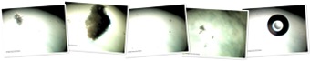
The photographs below represent a 5 day progression study of the lesion contents in a Petri Dish with nutrient agar at 100x:
Day 1

Day 2
Day 3
Day 5
How Morgellons Is Formed
IDENTIFICATION and PROCESS BEHIND FORMATION OF THE MORGELLONS SPHERES FROM FIBERS
These photos show how certain fibers bend to form the circular vesicles:
![[image]](https://i0.wp.com/my-stuff-dot-com.com/LB/Clathrin%20Coated%20Vesicles.jpg)
A diagram from the NIH:
http://www.ncbi.nlm.nih.gov/bookshelf/br….figure&id=A4895

"This diagram from this website shows how the Morgellons fungi are most likely formed from our ‘fibers’:
http://mycorhizes.com/Aarchaeosporales.html

"Saccule sporifPre et spore d’Acaulospora rehmii
Sporiferous saccule and spore of Acaulospora rehmii
or exhibit a spore dimorphism with the combination of acaulosporoVd and glomoVd types"
![[image]](https://i0.wp.com/mycorhizes.com/images/classificationShema12.jpg)
![[image]](https://i0.wp.com/mycorhizes.com/images/classificationPhoto16.jpg)
"Spore of the glomoVd type (Glomus mosseae)"
"The organisms are characterise by a distinc molecular signature at the level of the SSU rRNA gene “YCTATCYKYCTGGTGAKRCG” corresponding to the homolog position 691 of the Jo1353 sequence of Saccharomyces cerevisiae and specific to the taxon.
Families: Archaeosporaceae (Apendicispora, Archaeospora, Intraspora) and Geosiphonaceae (Geosiphon)"
![[image]](https://i0.wp.com/mycorhizes.com/images/glomeromycota-classification-archaeo_000.jpg)
This is also the genus and species of the fungi believed to be involved in Morgellons, photo comparisons to follow.
Morgellons Is Partially the Frog Fungus
Morgellons Is Partially the Frog Fungus Batrachochytrium dendrobatidis or Chytrid
I’m looking for Stock Photos of Batrachochytrium dendrobatidis on the Web to see if I can match them with my microscopic photos of cultured human Morgellons samples.
http://www.esf.edu/efb/brunner/research.htm

"Development of Batrachochytrium dendrobatidis zoospores."
The name of the photograph above is "ChytridDevelopment.png", this suggests that this is how the Chytrid species develops overall, more research is required at this time.
Below is a comparison from human ear lesion, cultured in nutrient agar at 30 days growth at 100x:

I’m noticing that there aren’t too many good microscopic stock photos, but this one above is a good example to give us an indication that Batrachochytrium dendrobatidis or a Chytrid fungus species is most likely involved as one of the mystery Morgellons fungi.
References:
http://www.sciencedaily.com/releases/2008/08/080812135654.htm
Morgellons Is Zoonotic
Frog / Koala Disease Related to Morgellons
The first years in my home in Atlanta, I had so many frogs around it in the summertime, it was so loud and eerie sounding, like a bad Alfred Hitchcock frog movie. Then the past 2 summers not a single frog, it was very quiet – I have to say, this is even more eerie! There had always been frogs around my house – where did all the frogs go?
http://jcm.asm.org/cgi/reprint/37/7/2378.pdf
"Chlamydia pneumoniae in a Free-Ranging Giant Barred Frog (Mixophyes iteratus) from Australia
The koala biovar of Chlamydia pneumoniae was identified in lung tissue from a sick, free-ranging giant barred frog (Mixophyes iteratus) by using electron microscopy, C. pneumoniae-specific fluorescent-antibody staining, cell culture, and sequencing of the ompA, ompB and 16S rRNA genes. This is the first report of a chlamydial strain infecting both a homeotherm and a poikilotherm and only the fourth host (in addition to humans, koalas, and horses) to be naturally infected with this species of Chlamydia. The frog had severe, chronic, mononuclear pneumonia and nonregenerative anemia and pancytopenia.
Chlamydia infections have been reported previously in captive amphibians, causing moderate to high mortality rates in various species…
There have also been a few case reports of Chlamydia infections in captive and wild reptiles. In all cases, however, the chlamydial species was either unknown or assumed to be C. psittaci. Here we report the first isolation of Chlamydia from a frog in Australia and demonstrate that it is identical to the C. pneumoniae strain that infects koalas."
I cannot link the photos in the .pdf file but look at the photograph in Figure 4 taken at 700nm, example "I", what they are calling example "condensed elementary bodies"; is very similar to a photograph that I took that I called "Mr. Brown Eye" cultured from my ear, 28 days growth, at 450x:

"The chlamydial particles included dense elementary bodies (314 6 39 nm in diameter [mean 6 standard deviation; n 5 12]), intermediate bodies (371 6 44 nm [n 5 12]), and numerous dividing reticulate bodies (537 6 130 nm [n 5 12]) (Fig. 4). The round elementary bodies had eccentric nuclei and a narrow or nonexistent periplasmic space. Mitochondria were not associated with the inclusion membrane. These ultrastructural characteristics are consistent with those of C. pneumoniae (14).
Several studies have reported the very high genetic similarity of human C. pneumoniae strains (5) and also the clonality of koala C. pneumoniae strains (24), suggesting a possible recent divergence of these biovars. It is possible that the infection of a wild frog described in this report was an isolated incident, or alternatively, increased testing may show that amphibians are commonly infected with C. pneumoniae and that they are a natural reservoir for this species."
We need to look at the various animals that are becoming sick in nature at this time to see if there is a correlation between their disease and ours, one inclusive of the bats, bees, frogs, and we need to especially look at what has happened to the koala and frogs with this Chlamydia aspect.
Once again, we have GIANT cells involved in our disease, look at "Mr. Brown Eye"; in the photo below, now at 100x, he’s bulging out of the frame! I have shown you thousands of photos of GIANT spheres taken at 100x.

The cell infecting the koala and frog are also GIANT C. pneumonia cells called "large inclusions"
Koala Biovar of Chlamydia pneumoniae Infects Human and Koala Monocytes and Induces Increased Uptake of Lipids In Vitro
http://iai.asm.org/cgi/content/abstract/69/12/7894
"We examined the ability of the koala biovar of Chlamydia pneumoniae to infect both Hep-2 cells and human monocytes and the effect of infection on the formation of foam cells. The koala biovar produced large inclusions in both human and koala monocytes and in Hep-2 cells."
It looks highly probable that those of us with evident Morgellons have a disease similar to what is eliminating the frog and koala populations. We also need to look at the diminishing butterfly, bees, amphibians, and bats.
Morgellons ‘Fibers’ Turn Into Spheres
Clathrin Coated Vesicles
Formed from Fibers
While studying this Chlamydia .pdf:
http://www.chlamydiae.com/docs/biology/biol_entry.asp
it opened a lot of doors for me – we now have names for some of the artifacts we’ve been seeing and proves some of the theory that we have proposed is most likely happening, such as; we have HeLa cells, photos to show this aspect later.
Figure 2.
"Fig 2. A Chlamydia trachomatis elementary body (EB) in the process of entry into a HeLa cell. Tannic acid stained to enhance visualisation of chlathrin. Note the clearly demarcated outer envelope of the EB and the surrounding membrane (m) of the vacuole. Also note how small the clathrin coated pit (ccp) is compared to the chlamydial endosome. The bar represents 0.1 microns."
We now have a name for our ‘carbon-looking balls’:

The ‘balls’ that are inside of the fungus gnat larvae @100x:

The same ones inside of our nematodes, Urine at 600x:

They are called a clathrin coated pit, labeled "ccp" in the diagram above.
If we research the clathrin coated pit, we see how certain fibers form these spheres:
"Reconstitution of Clathrin-coated
Pit Budding from Plasma Membranes"
http://jcb.rupress.org/cgi/reprint/114/5/881.pdf
These photos show how certain fibers bend to form the circular vesicles:

A diagram from the NIH at:
http://www.ncbi.nlm.nih.gov/bookshelf/br.fcgi?book=mcb&part=A4895&rendertype=figure&id=A4895
Our Morgellons Spheres
An Abiogenic Life Form?
When I take regular debris out of my lesion – the spheres start out looking like what I have described as a ‘biofilm’ – they are translucent in color and circular, such as this photo below, a little more translucent than this photo:

With saliva and one other time when I had a phosphate remedy on my ear that also entered the Petri Dish, they showed up as white circular growths that I described as looking like Pseudomonas aeruginosa in a Petri Dish when comparing it to online stock photos.
These dishes contain all white circular growths and NONE of the others, such as the greens, browns, blacks, greys, orange, white fuzzy, etc. growths that we’re seeing in most. Eventually – as noted in certain experiments – these ‘others’ will have appeared:
![[image]](https://i0.wp.com/my-stuff-dot-com.com/LB/yeast%20integrating%20plasmid.jpg)
Our yeast/fungus has a biofilm involved, in the petri dishes it is a light to mustard yellow in color, you will see this on the bottom side of the dish in a couple of weeks time.
We’re looking for a fungus/yeast that starts out as translucent (like a biofilm), then turns to white circular cocci-shaped growth and then produces a yellowish biofilm as a by-product in a petri dish and I believe this same process is happening under our skin.
What’s been so confusing, and this is where Frito can help us by studying the progressions, is… that even though we’re starting with one sphere – there seems to be approximately 5 or more different characteristics that they end up with. They all don’t perform the same way. Some of them stay spheres and allow the ‘spaceship’ (polyhedron) to land on them, some of them explode into a white fungus, some of them explode into a black/white fungus, some of them have ‘fibers’ laying on them or nearby in which they are appear to be quorum sensing with, some of them morph into ‘other’ different-looking spheres, and they reproduce in many different ways.
What I have described in the past as the ‘biofilm’ IS the main sphere. Out of the biofilm all things are created. It’s as if we have a multi-faceted combination of events occurring out of our spheres – such as a possible baculoviral, protozoa-like artifacts, our mystery fibers, some sprouting into fungi, some are occluding, others not, some are budding, others not… etc.
The main sphere is like a biofilm, I want to say it is more of a yeast than fungus – the main ‘yeast’ sphere LATER creates a fungi or mold from inside of it – it appears that our sphere has properties that it can incorporate many other ‘things’ that come along into its ‘system’.
This could explain why some of us have different symptoms… we all have this ‘Master Sphere’ in common and what it does is responsible for the three things we all have in common – the specks/spheres, biofilm and fibers, the cause of Morgellons – however, not all of us have been subjected to the same things in our environments and lives.
Our sphere is like a vacuum cleaner sphere that robs, sucks in, takes in, incorporates, morphing and creating… more and more variables within it. It’s the ‘house’ for everything.
A TWO-PART SYSTEM
When you spit into a Petri Dish, and look microscopically – what do you see? Spheres? No, we see the micelles – that we are all starting to capture in our photographs… but, we don’t see them being large like our big sphere – what do they turn into? I believe that our Master Sphere has to take on so much liquid, undergo chemical processes, temperature, time, and grow to a certain size before they start budding, dividing, etc.
You initially see ‘fibers’ and ‘specks’… these specks are not spheres. There are some very small ‘micelles’ seen though, I believe I have shown that these eventually turn into our Master Spheres in observing them in the red wine culture experiment on my blog site.
Here’s a photo of Jeany’s saliva one day after, 100x:
![[image]](https://i0.wp.com/my-stuff-dot-com.com/LB/11_09_221.jpg)
And then on the 3rd day… as Frito said, look at the size these things can get to be in a hurry!…
![[image]](https://i0.wp.com/my-stuff-dot-com.com/LB/11_11_111.jpg)
Most of the fibers are gone, there was just one odd looking fiber left on day 3…
We’re looking for an invention that starts with a ‘fiber’ (worm micelle) that can ‘sprout’/create an adaptable, morphing ‘Master Sphere’. I believe it creates these spheres on a very small scale and start out as what is known as a sphere micelle. I don’t believe that the fungus/mold that we’re looking for is entirely a known fungus, it’s a hybrid that’s been manipulated to be created from out of the sphere and the ‘fiber’ was its origin. Our Master Sphere is most likely something else… a yeast, a housing system, a larger micelle, that can hold fungi, mold, yeast, viruses, bacteria, and ‘others’.
I do believe from what we’re seeing, a ‘fiber’ that creates this Master Sphere is in certain red wine-making, cotton production and paper-making processes, it is in some areas of the country’s water remediation processing… that it is quite well-known and considered safe – if it is, then – why can’t we easily identify it?
A flowchart of what’s happening might look like:
Fiber –> Micelle –> Biofilm –> Yeast Cell ? –> Fungus/Mold/Others
So What Are Our Spheres?
I have called our spheres many different names – Lysosomes, micelles, vesicles, Hulle cells, fungal spores, Carnicom is finding hemoglobin…
This has been very puzzling to me – Why? Because what I proposed above defies the laws of nature. The laws of nature are…
Biogenesis
(1) The process in which life forms arise from similar life forms.
(2) It asserts that living things can only be produced by another living thing, and not by a non-living thing.
Spontaneous generation
The previously popular notion that living organisms arise or develop from nonliving matter.
Abiogenesis
"In the natural sciences, abiogenesis, or "chemical evolution", is the study of how life on Earth could have arisen from inanimate matter. It should not be confused with evolution, which is the study of how groups of living things change over time." [1]
What I am theorizing, based on what I am witnessing in a Petri Dish with a microscope, is that Morgellons is utilizing Abiogenesis or a form of it, or some similar new, unheard-of science in our disease process.
Common sense tells us that if we have a chicken egg, and we hatch it – we’re going to get a chick – every time! We’re not going to get 5 different possibilities that might come out…! (I am still not certain that some of us might not have more than one sphere involved.)
What our main sphere is – is of utmost importance in understanding our disease, only then will we be able to get a better handle on what has happened to us, what’s going on and how to manage it.
References:
The Morgellons "Spaceships"
Triclinic Crystal Clusters?…
Here is a stock photo of a triclinic crystal cluster:

I’ve written quite a few posts about the Morgellons "spaceships", not knowing what I am seeing – I had to call them something… they look like something out of a "Star Wars" movie, they look like they’re flying in the Petri Dish, and they seem to appear and disappear as if, they have movement.
All photos are from human lesion samples, 100x:

They appear metallic in nature, as if this is a charcoal pencil drawing with the added green color. I have stated that I believe they are carbon-like from their appearance.
I have not studied them continuously, day to day… I photograph every few days, so – I have missed a lot of how they are formed and what they are doing in the scheme of things. They don’t appear in the Petri Dish until approximately Day 20 or so… they are formed by the sphere coming into direct or quorum sensing contact with the hyphae of the fungus or from a ‘fiber’.


They first appear as ‘bumps’ on the main ‘big sphere’ believed to be a fungal spore? But, not all of the spheres/spores do this. Which might indicate that they are a part of the inside chemistry of the fungus?

They next appear to be resting on the sphere:


I am not sure, but I believe this is a ‘spaceship’ being born from inside the sphere when it buds:

I have seen evidence of where they possibly could be being ‘born’ from the fibers themselves:

We see them intermixed with the hyphae of the fungus or scattered about randomly. If they are conceived inside the spheres to me, this indicates movement.:

Then they could be in this photo of where a sphere has exploded them out?:

They are never the same in appearance, but have some features in common.
The ‘Mother Spaceships’ can reproduce ‘Baby Spaceships’…


We will soon look at how these Morgellons "Spaceships" might be related to triclinic crystal clusters.
-
Recent
- Morgellons Artifacts Found in Rainwater – Part 1
- Morgellons Researchers
- Morgellons Disease Sufferers Growing Weary in the Wait
- Morgellons Progression Study
- How Morgellons Is Formed
- Morgellons Is Partially the Frog Fungus
- Morgellons Is Zoonotic
- Morgellons ‘Fibers’ Turn Into Spheres
- Our Morgellons Spheres
- The Morgellons "Spaceships"
- A Lot of Fiber in our Diet?
- Cigarette Filter Fiber Study
-
Links
-
Archives
- April 2010 (3)
- March 2010 (5)
- November 2009 (6)
- October 2009 (13)
- September 2009 (4)
- June 2009 (13)
-
Categories
-
RSS
Entries RSS
Comments RSS
